The SMS offers high performance Raman microspectroscopy with specifications comparable to high-end benchtop systems, but with unrivaled flexibility.
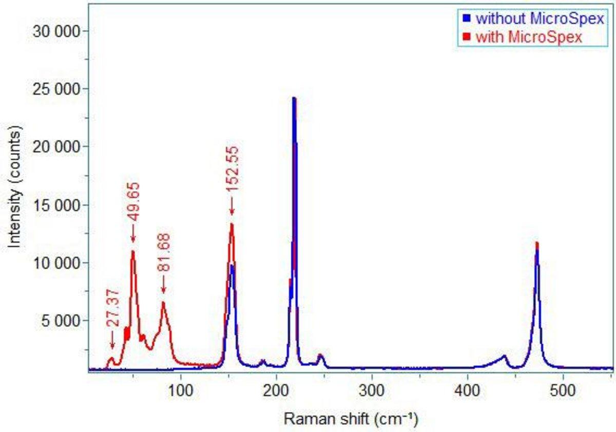
Raman spectra of sulfur (red) measured on SMS system.
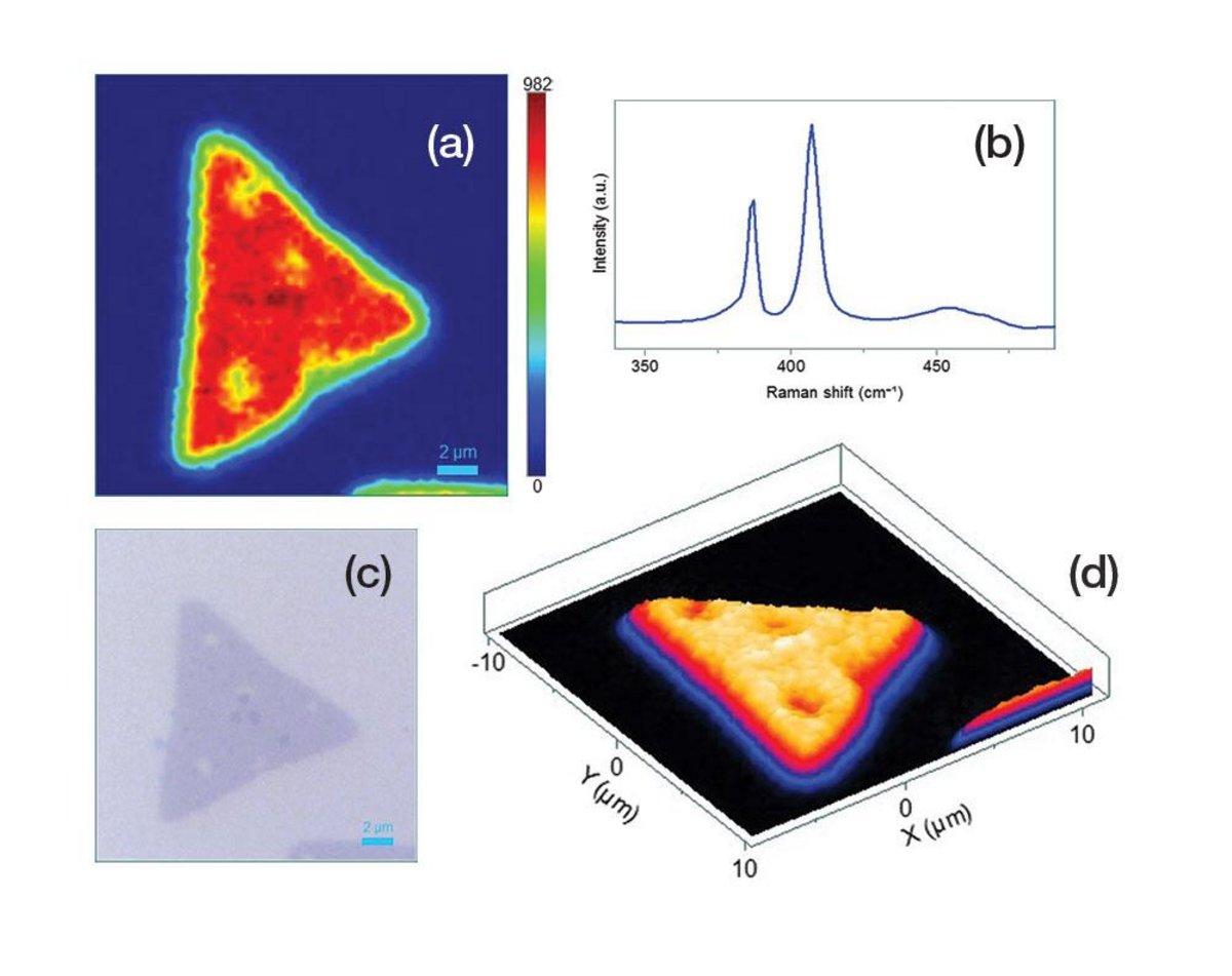
Raman characterization of a monolayer MoS2 using 532 nm laser line (objective = 100x, spatial resolution = 0.3 μm). (a) 2D image of Raman map of A1g peak intensity. (b) A Raman spectrum showing sharp and intense E1 and A1g peaks of MoS2 sample. (c) Optical image of the MoS2 flake. (d) 3D image of Raman map of A1g peak intensity.
| Spectrometer and Detectors | |||||
|---|---|---|---|---|---|
| Spectrometers | MicroHR | iHR320 | iHR550 | ||
| Excitation Lasers | 532 nm, 633 nm, 785 nm | ||||
| Spectral Range (cm-1) – Free space | 80 – 9500 (532 nm), 80 – 6500 (633 nm), 80 – 3400 (785 nm) | ||||
| Spectral Range (cm-1) – Fiber | 150 – 9500 (532 nm), 150 – 6500 (633 nm), 150 – 3400 (785 nm) | ||||
| Recommended Gratings | 1800 g/mm, 1200 g/mm, 600 g/mm | ||||
| Spectral Resolution1 (cm-1/ pixel) | 405 nm | 5.12 | 2.34 | 1.37 | |
| 532 nm | 2.53 | 1.22 | 0.73 | ||
| 633 nm | 1.54 | 0.78 | 0.48 | ||
| 785 nm | 0.70 | 0.40 | 0.26 | ||
| Microscope | |||||
|---|---|---|---|---|---|
| Microscope Objectives | Magnification | 10X | 50X | 100X | |
| Spot Size (fiber-coupled) | < 50 μm | < 12 μm | < 6 μm | ||
| Spot Size (free space-coupled) | < 10 μm | < 5 μm | < 2 μm | ||
| Sample Stage | XYZ (Manual and motorized options available) – 75 x 50 mm; 100 x 100 mm; 150 x 150 mm; 300 x 300 mm | ||||
| Vision Camera | Software controlled vision camera included | ||||
1 For 1800 g/mm grating and 26 μm pixel CCD
Do you have any questions or requests? Use this form to contact our specialists.

